The human skull represents one of the most complex and fascinating structures in the human body. Composed of multiple interconnected bones, the skull serves as both a protective fortress for our brain and the structural foundation for our facial features. Understanding skull anatomy, particularly the distinction between the neurocranium and viscerocranium, provides valuable insights into human evolution, medical diagnosis, and surgical procedures.
What is the Human Skull?
The human skull is a bony structure that forms the head’s framework, consisting of 22 individual bones that fuse together during development. This remarkable structure weighs approximately 2.3 kilograms (5 pounds) in adults and serves multiple critical functions beyond simple protection. According to research published in medical journals and referenced by Wikipedia, the skull’s primary purposes include protecting the brain, housing sensory organs, and providing attachment points for facial muscles.
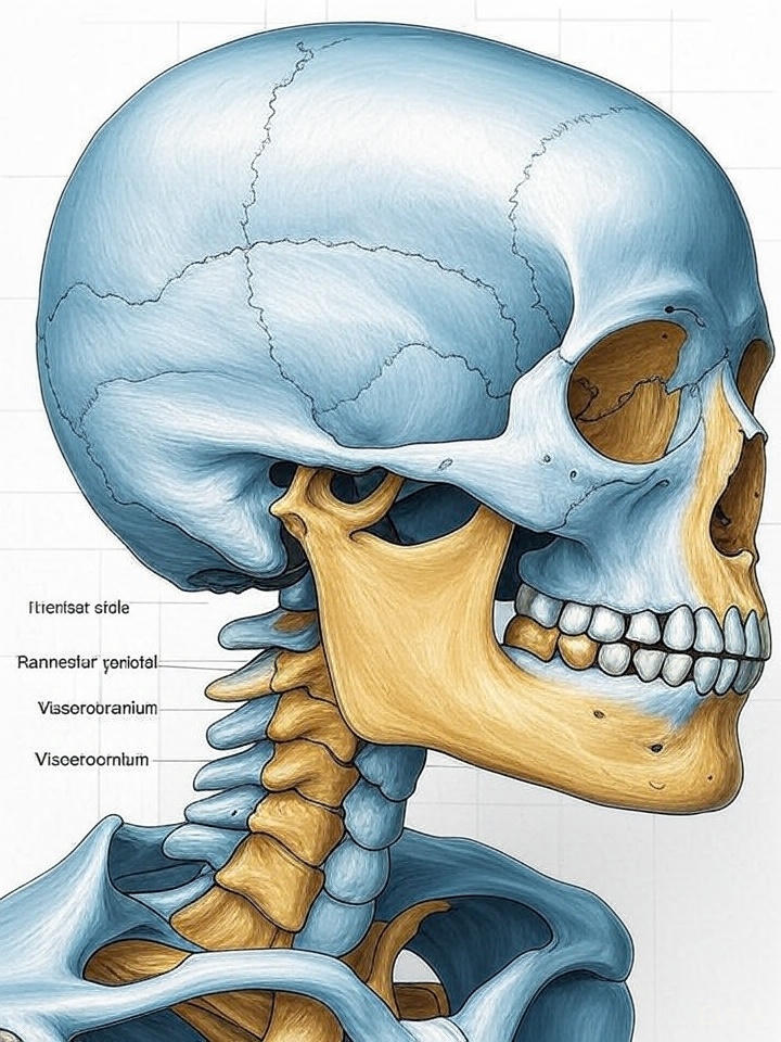
The skull’s complexity becomes apparent when examining its two main divisions: the neurocranium (brain case) and the viscerocranium (facial skeleton). These two components work together to create the distinctive human head shape while accommodating various biological functions.
Understanding the Neurocranium: The Brain’s Protective Shell
The neurocranium, also known as the cranial vault or brain case, forms the protective housing for the brain and brain stem. This structure consists of eight cranial bones that create a nearly spherical cavity called the cranial cavity. The neurocranium’s primary function involves protecting the delicate neural tissue while providing structural support for the head.
Components of the Neurocranium
The neurocranium comprises several distinct cranial bones, each contributing to the overall protective structure:
Frontal Bone: Located at the front of the skull, the frontal bone forms the forehead and the upper portion of the eye sockets. This bone contains the frontal sinuses, air-filled cavities that help reduce the skull’s weight while maintaining strength.
Parietal Bones: These paired bones form the sides and roof of the skull. The parietal bones are among the largest cranial bones and play a crucial role in protecting the brain’s parietal lobes, which are responsible for processing sensory information.
Temporal Bones: Positioned on either side of the skull, the temporal bones house the ear structures and contain important foramina (openings) for nerves and blood vessels. These bones also feature the mastoid process, where neck muscles attach.
Occipital Bone: Found at the back and base of the skull, the occipital bone contains the foramen magnum, the large opening through which the spinal cord connects to the brain stem.
Sphenoid Bone: This complex, butterfly-shaped bone sits at the skull’s base and houses the pituitary gland in a depression called the sella turcica. The sphenoid bone also forms part of the eye socket walls.
Ethmoid Bone: Located between the nasal cavity and the brain, the ethmoid bone contributes to the nasal septum and contains numerous air cells that form part of the paranasal sinus system.
Neurocranium Development and Clinical Significance
The neurocranium develops through a process called intramembranous ossification, where bone tissue forms directly from mesenchymal tissue. This development process is crucial for allowing brain growth during infancy and childhood. Medical professionals, as reported by institutions like Mayo Clinic, monitor neurocranium development to detect potential abnormalities such as craniosynostosis, where skull sutures fuse prematurely.
Understanding neurocranium anatomy proves essential for various medical specialties, including neurosurgery, radiology, and emergency medicine. Healthcare providers must thoroughly understand these structures when treating head injuries, planning surgical procedures, or interpreting diagnostic imaging.
Exploring the Viscerocranium: The Facial Skeleton
The viscerocranium, commonly referred to as the facial skeleton, comprises 14 bones that form the face’s structural framework. Unlike the neurocranium, which primarily serves protective functions, the viscerocranium supports various physiological processes including breathing, eating, speaking, and facial expression.
Major Components of the Viscerocranium
The facial skeleton includes several key bones, each serving specific functions:
Maxilla Bones: These paired bones form the upper jaw and contain the upper teeth. The maxillae also contribute to the nasal cavity floor and the hard palate formation. Additionally, these bones house the maxillary sinuses, the largest paranasal sinuses.
Mandible: The mandible represents the largest and strongest facial bone, forming the lower jaw. This U-shaped bone holds the lower teeth and provides attachment points for powerful chewing muscles. The mandible is the only movable skull bone, enabling essential functions like speaking and eating.
Nasal Bones: These small, paired bones form the bridge of the nose and support the cartilaginous portion of the nasal structure.
Zygomatic Bones: Commonly known as the cheekbones, these bones form the prominence of the cheeks and contribute to the eye socket walls.
Palatine Bones: These L-shaped bones help form the hard palate and nasal cavity walls, playing crucial roles in separating the oral and nasal cavities.
Lacrimal Bones: The smallest facial bones, located in the medial wall of each eye socket, contain grooves for tear duct structures.
Functional Importance of Viscerocranium
The viscerocranium’s design reflects its multiple functional requirements. Research published by medical institutions and referenced in Forbes Health articles emphasizes how facial bone structure directly impacts breathing efficiency, speech clarity, and overall quality of life.
The upper respiratory tract, which includes structures supported by viscerocranium bones, connects to the lower respiratory system. Understanding this connection becomes particularly important when examining conditions affecting respiratory health, as detailed in comprehensive medical resources about lower respiratory tract infections.
Skull Anatomy in Medical Practice
Medical professionals rely heavily on detailed skull anatomy knowledge for various diagnostic and therapeutic procedures. Radiologists interpret CT scans and MRIs by identifying specific cranial bones and their normal anatomical relationships. Surgeons planning procedures must understand the precise locations of vital structures relative to skull landmarks.
Diagnostic Applications
Modern medical imaging techniques allow healthcare providers to examine skull anatomy with unprecedented detail. Computed tomography (CT) scans provide excellent visualization of bone structures, while magnetic resonance imaging (MRI) offers superior soft tissue contrast. These imaging modalities help diagnose conditions ranging from traumatic injuries to developmental abnormalities.
Medical professionals use specific anatomical landmarks on cranial bones to guide procedures and interpret findings. For example, the external auditory meatus in the temporal bone serves as a reference point for various measurements and surgical approaches.
Surgical Considerations
Neurosurgical procedures require intimate knowledge of skull anatomy, particularly the relationship between cranial bones and underlying brain structures. Surgeons must understand the thickness variations of different skull regions, the locations of major blood vessels, and the positions of critical nerve pathways.
Oral and maxillofacial surgeons working with viscerocranium structures must appreciate the complex relationships between facial bones, teeth, and surrounding soft tissues. Procedures involving the mandible, for instance, require understanding of muscle attachments, nerve pathways, and blood supply patterns.
Evolutionary Perspective on Skull Development
The human skull’s current form represents millions of years of evolutionary adaptation. Anthropologists and evolutionary biologists study skull anatomy to understand human development and our relationship to other species. The expansion of the neurocranium relative to the viscerocranium reflects the significant growth of human brain capacity throughout evolution.
Comparative Anatomy
When compared to other primates, the human skull shows several distinctive features. Our neurocranium is proportionally larger, reflecting increased brain size, while our viscerocranium is relatively reduced, particularly in the jaw region. These changes correlate with dietary shifts and the development of tool use in human ancestors.
The positioning of the foramen magnum in the occipital bone also differs significantly between humans and other primates. In humans, this opening sits more anteriorly, reflecting our upright posture and bipedal locomotion.
Clinical Conditions Affecting Skull Anatomy
Various medical conditions can affect both the neurocranium and viscerocranium, ranging from congenital abnormalities to acquired diseases. Understanding these conditions requires thorough knowledge of normal skull anatomy and development patterns.
Congenital Abnormalities
Craniosynostosis represents one of the most common congenital skull abnormalities, occurring when cranial sutures fuse prematurely. This condition can affect neurocranium growth and may require surgical intervention to allow proper brain development.
Cleft lip and palate conditions affect viscerocranium development, specifically involving the maxilla and palatine bones. These conditions require multidisciplinary treatment approaches involving surgeons, dentists, and speech therapists.
Traumatic Injuries
Skull fractures can affect either the neurocranium or viscerocranium, depending on the location and mechanism of injury. Healthcare providers classify these fractures based on their anatomical location and potential complications.
Mandible fractures are among the most common facial bone injuries, often resulting from motor vehicle accidents or interpersonal violence. Treatment requires understanding of mandibular anatomy, including muscle attachments and dental relationships.
Advanced Imaging and Skull Anatomy
Modern medical technology has revolutionized our ability to visualize and study skull anatomy. Three-dimensional reconstruction techniques allow clinicians to examine complex anatomical relationships and plan surgical procedures with unprecedented precision.
CT and MRI Applications
High-resolution CT scans provide excellent detail of cranial bones and can detect subtle fractures or developmental abnormalities. Radiologists use specific protocols optimized for skull imaging, adjusting parameters based on the clinical question and patient factors.
MRI technology excels at imaging soft tissues within and around the skull, including brain tissue, blood vessels, and nerves. Advanced MRI techniques can even visualize cranial nerve pathways and their relationships to surrounding bone structures.
3D Reconstruction Technology
Computer-assisted three-dimensional reconstruction allows surgeons to practice procedures on virtual models before operating on patients. This technology is particularly valuable for complex reconstructive procedures involving both neurocranium and viscerocranium components.
Medical education also benefits from 3D visualization technologies, allowing students to explore skull anatomy in ways impossible with traditional cadaveric specimens or textbook illustrations.
Future Directions in Skull Anatomy Research
Ongoing research continues to expand our understanding of skull anatomy and its clinical applications. Areas of active investigation include the development of biocompatible materials for skull reconstruction, improved imaging techniques for early disease detection, and better understanding of genetic factors controlling skull development.
Regenerative Medicine Applications
Scientists are exploring tissue engineering approaches to reconstruct damaged skull bones using patient-derived stem cells and biodegradable scaffolds. These techniques could revolutionize treatment of large skull defects resulting from trauma, infection, or tumor removal.
Genetic Research
Advances in genetic research have identified numerous genes controlling skull development and growth. Understanding these genetic mechanisms may lead to improved treatments for congenital abnormalities and better prediction of developmental problems.
Conclusion
The human skull’s intricate anatomy reflects millions of years of evolutionary adaptation and serves multiple critical functions in modern humans. Understanding the distinction between neurocranium and viscerocranium components provides essential knowledge for medical professionals, researchers, and students alike.
From the protective neurocranium housing our most vital organ to the complex viscerocranium enabling communication and nutrition, each component of skull anatomy contributes to human survival and quality of life. As medical technology continues advancing, our appreciation for these remarkable structures grows, leading to improved diagnostic techniques and treatment options.
The study of skull anatomy connects multiple medical specialties and research fields, from basic anatomical education to cutting-edge regenerative medicine applications. Whether examining cranial bones for diagnostic purposes or planning complex surgical procedures, thorough understanding of skull anatomy remains fundamental to modern medical practice.
As we continue exploring the complexities of human anatomy, the skull serves as a perfect example of form following function, demonstrating how evolutionary pressures shaped the remarkable structure that defines our species’ unique capabilities and distinguishes us within the animal kingdom.
Leave a Reply