Anatomy of the Skull: Introduction to the Cranium & Viscerocranium
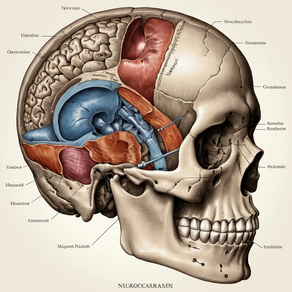
The skull is a complex, protective shell housing the brain and the entrance to vital systems like the respiratory and digestive tracts. It’s split into two major divisions: the neurocranium (braincase) and viscerocranium (facial skeleton). A solid understanding of the bones of the cranium and the intricacies of skull osteology is essential for medical, dental, and anatomical disciplines.
→ For foundational definitions, see Wikipedia: Skull.
Divisions of the Skull
The skull is made up of 22 bones, with the neurocranium forming the protective case around the brain and the viscerocranium shaping the facial skeleton. The neurocranium includes 8 bones (frontal, 2 parietal, 2 temporal, occipital, sphenoid, and ethmoid), while the viscerocranium contributes the rest.
To understand how cranial nerves traverse this structure, see our in-depth post on Cranial Nerves Explained.
Overview: Paired vs Unpaired Cranial Bones
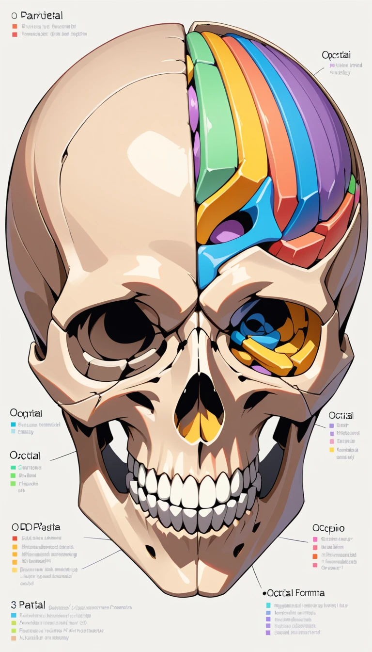
The bones of the cranium are categorized as either paired (parietal, temporal) or unpaired (frontal, occipital, sphenoid, ethmoid). Each bone plays a structural and functional role in forming the cranial vault and base.
Frontal Bone: Anatomy & Articulations
The frontal bone forms the forehead, part of the orbital roof, and nasal cavity. It includes:
Squamous part (forehead)
Orbital part (superior orbit)
Nasal part (articulates with nasal bones)
The supraorbital margin contains the supraorbital notch or foramen, a key neurovascular landmark.
Parietal Bone & Calvaria
The parietal bones form the roof and sides of the cranial vault. They meet at the sagittal suture and articulate anteriorly with the frontal bone, forming the bregma. The calvaria (skull cap) includes the parietal bones and contributes to the vertex and lambda points—landmarks used in craniometric analysis.
Temporal Bone: Squamous, Tympanic, Petromastoid Parts
This bone is complex, forming parts of the ear canal, middle ear, and base of the skull. Its parts include:
Squamous (thin upper plate)
Tympanic (surrounds external acoustic meatus)
Petromastoid (dense, protects cochlea and semicircular canals)
The temporal bone also contains the mandibular fossa—forming the temporomandibular joint (TMJ), an essential structure in jaw articulation.
Occipital Bone: Basilar, Condylar & Foramen Magnum
Located at the skull’s posterior, the occipital bone houses the foramen magnum, through which the brainstem passes. The condyles articulate with the first cervical vertebra (atlas), while the basilar part connects with the sphenoid bone.
Sphenoid Bone: Body, Wings & Sinuses
The sphenoid sits at the base of the cranium and is called the “keystone” of the skull. It has:
A central body (housing the sphenoidal sinus)
Greater and lesser wings
Multiple foramina: foramen rotundum, ovale, and spinosum, transmitting cranial nerves and vessels
Ethmoid Bone: Structure & Location
The ethmoid bone contributes to the medial wall of the orbit, the nasal cavity, and part of the anterior cranial base. It contains the cribriform plate (for CN I) and supports paranasal sinus drainage into the nasal passages.
Calvaria & Cranial Base: Superior & Basal Anatomy
The calvaria (skull cap) forms the top of the cranial vault. Key landmarks include:
Vertex (highest point)
Bregma (junction of coronal and sagittal sutures)
Lambda (intersection of sagittal and lambdoid sutures)
The cranial base is divided into three fossae:Anterior cranial fossa: holds frontal lobes
Middle cranial fossa: houses temporal lobes and pituitary gland
Posterior cranial fossa: accommodates the cerebellum and brainstem
Growth & Closure: Sutures & Fontanelle Timing
Cranial sutures allow skull expansion during infancy. They include:
Coronal suture
Sagittal suture
Lambdoid suture
Fontanelles (soft spots) facilitate birth and brain growth:Anterior fontanelle (bregma): closes by 18–24 months
Posterior fontanelle (lambda): closes by 2–3 months
These are critical clinical landmarks when assessing developmental anomalies.
Cranial Foramina Mapping
The skull base features multiple foramina that transmit nerves and blood vessels.
Notable examples:
Optic canal (CN II)
Superior orbital fissure (CN III, IV, V1, VI)
Foramen ovale (CN V3)
Jugular foramen (CN IX, X, XI)
Hypoglossal canal (CN XII)
Also important is the middle meningeal artery which enters via the foramen spinosum—a common site in epidural hematomas.
To explore these in detail, visit Cranial Nerves Explained.
Cranial Ossification Centers & Vault Growth
Cranial bones form via two main mechanisms:
Intramembranous ossification: flat bones like the frontal and parietal bones
Endochondral ossification: base of the skull (sphenoid, occipital)
Cranial ossification centers appear during embryogenesis. Their timing and fusion determine skull shape and are critical in diagnosing conditions like craniosynostosis.
Vault growth continues into adolescence, with sutures gradually fusing into adulthood.
Clinical Cases & Surgical Considerations
Understanding cranial osteology aids in diagnosing:
Skull fractures: Depressed, basilar, or growing fractures in children
Sinusitis: Involvement of ethmoid or maxillary sinuses
TMJ disorders: Dysfunction of the temporomandibular joint can affect chewing and speech
CSF leaks: Due to fractures or surgical complications
Surgical interventions often involve access to the skull base, such as transsphenoidal surgery for pituitary tumors. Understanding anatomical landmarks prevents complications.
For practical insights, see neurosurgical discussions on platforms like Forbes Health or Mayo Clinic.
Human Skull Evolution
The structure of the braincase has evolved over millennia. Early hominins like Homo erectus had smaller, elongated skulls with robust brow ridges. Homo sapiens developed a rounder neurocranium and vertical forehead—supporting increased brain volume.
This shift in cranial morphology is studied extensively in anthropology and supports theories of language, cognition, and tool use development.
Recap & Interactive Quiz
Quick summary of what we covered:
Eight bones form the neurocranium
Foramina transmit critical nerves and vessels
Sutures and fontanelles allow skull growth
Surgical access and developmental timing depend on precise anatomy
Quiz examples:
Which cranial nerve passes through the foramen rotundum?
What forms the anterior fontanelle?
Which bones form the calvaria?
Leave a Reply