
📋 Table of Contents
1. Introduction to Cranial Anatomy <a name=”introduction”></a>
1.1 What Is “Cranial” vs “Cranium”?
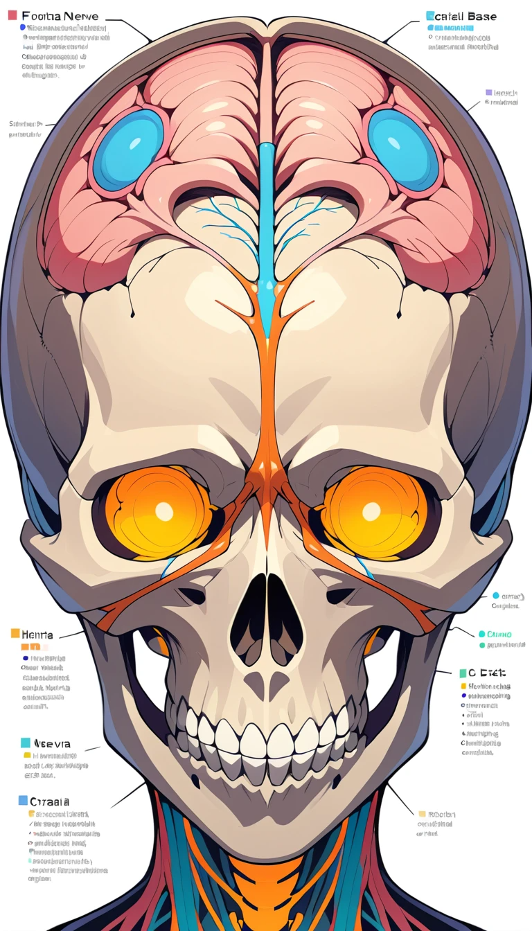
“Cranial” and “cranium” stem from the Greek kranion, meaning skull. In anatomy, “cranial” refers broadly to the head region, while the “cranium” specifically is the bony structure protecting the brain.
1.2 Etymology: Greek Roots
The term cranium derives from the Greek kranion: a foundational term that appears frequently in medical and anatomical language—think cranioplasty, craniosynostosis, or cranial nerves.
2. Skull (Cranium) Structure <a name=”skull-structure”></a>
2.1 Neurocranium vs Facial Skeleton
Neurocranium (braincase): Encloses and protects the brain.
Facial skeleton: Forms eyes, nose, jaw, and upper teeth.
Understanding neurocranium vs facial skeleton differences is key in anatomy, anthropology, and forensic science.
2.2 Cranial Bones: Overview & Types
The adult cranium features eight bones—frontal, two parietal, two temporal, occipital, sphenoid, and ethmoid—that fuse through complex sutures.
2.3 Sutures & Skull Ossification
Major sutures include the coronal, sagittal, and lambdoid. Ossification timelines—like when the metopic suture closes—impact development and are crucial in pediatric evaluation.
3. Cranial Measurements and Indices <a name=”cranial-measurements”></a>
3.1 Cranial Index: Ratio & Norms
The cranial index (breadth/length × 100) informs classification:
Brachycephalic (≥ 81): wide skulls.
Dolichocephalic (≤ 75): elongated skulls.
3.2 Cranial Capacity: Braincase Volume
Cranial capacity is the cubic volume of the braincase—important in evolutionary anthropology and neurological study. Norms vary across populations.
3.3 Cranial Module: Dimensional Averages
The cranial module averages multiple linear skull measurements to assess overall size and shape—useful in forensic science and comparative anatomy.
4. Cranial Cavity & Foramina <a name=”cranial-cavity”></a>
4.1 The Cranial Cavity Explained
This is the enclosed space supporting and shielding the brain—divided into floors and vaults.
4.2 Foramina: Passageways & Functions
Tiny holes like the foramen magnum, optic canal, and jugular foramen transmit nerves and vessels:
Foramen magnum: spinal cord transition.
Optic canal: optic nerve & ophthalmic artery.
Jugular foramen: jugular vein, IX–XI cranial nerves.
4.3 Cranial Base (Cranial Floor)
The base articulates with facial bones and supports the brain. Its shape and angle matter in neurosurgery and paleontology.
5. Cranial Nerves Explained <a name=”cranial-nerves”></a>
Explore the twelve cranial nerve pairs—sensory, motor, and mixed—highlighting their clinical importance.
5.1 Overview: 12 Pairs
I (Olfactory)
II (Optic)
III–VI (Eye movement)
VII (Facial expression)
VIII (Hearing & balance)
IX–XII (Taste, swallowing, tongue movements)
5.2 Classification: Sensory, Motor, Mixed
Sensory: I, II, VIII
Motor: III, IV, VI, XI, XII
Mixed: V, VII, IX, X
5.3 Descriptions I–XII & Clinical Relevance
I (Olfactory): smell detection (anosmia concerns).
II (Optic): vision and visual fields.
III, IV, VI: ocular motor—palsy leads to strabismus or diplopia.
VIII: auditory assessments using tuning fork.
VII: facial symmetry tests; Bell’s palsy.
IX–X: gag reflex, vocal cords, heart rate.
XI (Accessory): SCM/trapezius strength.
XII (Hypoglossal): tongue movement (speech/swallow disorders).
For a deeper dive, check out Cranial Nerves Explained on my own site: “Cranial Nerves Explained”.
6. Cranial Functionality & Autonomic Interactions <a name=”cranial-functionality”></a>
6.1 Shielding & Housing the Brain
The neurocranium absorbs impacts and connects with meninges over the brain.
6.2 Role in Sensory and Motor Functions
Protects sensory organs, enables mastication, swallowing, facial expression, vision, and more.
6.3 Autonomic Nervous System Interface
Cranial nerve X (vagus) influences parasympathetic control of heart, lungs, and digestive organs—demonstrating integration with autonomic systems.
7. Clinical and Developmental Insights <a name=”clinical-insights”></a>
7.1 Common Conditions
Meningitis: inflammation of cranial meninges.
Trauma: fractures with hemorrhage in cranial cavities.
Craniosynostosis: early suture closure affecting brain growth.
7.2 Cranial Kinesis in Comparative Anatomy
Cranial kinesis—movement of skull bones separate from human ornithology—common in birds, reptiles, and fish. Comparative works highlight this trait’s adaptive function.
7.3 Measurement Uses in Anthropology & Medicine
Using cranial base foramina list, researchers trace evolutionary lineage. Radiologists apply cranial thickness measurements to evaluate trauma and developmental anomalies.
8. Conclusion & Applications <a name=”conclusion”></a>
Summary: Understanding cranium structure, nerve networks, measurements, and clinical relevance is essential in anatomy, medicine, and anthropology.
Applications:
- Medical education & neurology
- Clinical diagnosis (nerve palsy, trauma)
- Anthropology & comparative studiesNext Steps: Expand content with muscle, vasculature, and advanced imaging topics.
9. References & Further Reading <a name=”references”></a>
Wikipedia: Cranial nerves
Forbes Health: Brain Anatomy Guide
Your article: “Cranial Nerves Explained”
Leave a Reply