
3. The Cranial Cavity: What It Is & How Big It Gets 
3.1 What is the Cranial Cavity?
The cranial cavity—known in Latin as cavitas cranii—is the internal space made by the 8 neurocranium bones that houses the brain. It also contains the protective meninges and cerebrospinal fluid (CSF).
3.2 Average Cranial Capacity in Adults
In adult humans, cranial capacity typically ranges from 1,200 to 1,700 cm³, with average values around 1,400–1,500 cm³.
3.3 Why Volume Matters
The cranial cavity isn’t just a brain container. It’s a carefully engineered space that allows fluid flow, pressure equalization, and nerve protection. Any variation—such as swelling, bleeding, or compression—can result in serious complications.
4. Protective Functions of the Cranial Cavity
4.1 Brain Protection & Meninges
The brain is secured by three layers of meninges and bathed in cerebrospinal fluid, which cushions against shocks. These reduce trauma during impacts, while fractures can lead to dangerous bleeding or infection.
4.2 Cranial Nerves
Twelve pairs of cranial nerves pass through foramina in the neurocranium, controlling critical functions like smell, sight, chewing, swallowing, and facial movement.
4.3 Pituitary Gland Housing
The sella turcica, a saddle-like depression in the sphenoid bone, protects the pituitary gland, a hormone powerhouse regulating growth, stress response, and reproduction.
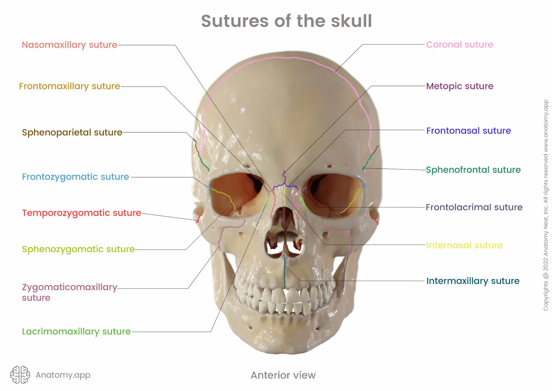
5. Skull Development: Sutures, Fontanelles & Ossification
5.1 Sutures & Fontanelles
Cranial sutures (coronal, sagittal, lambdoid, and squamous) are fibrous joints connecting skull bones. These remain open during infancy to allow brain growth and skull molding.
Fontanelles (soft spots):
Anterior: closes between 9 and 18 months.
Posterior: usually closes by 2–3 months.
Others (sphenoidal, mastoid): close by 6–18 months.
5.2 Ossification Patterns
Most cranial bones form via intramembranous ossification, where bone develops directly from tissue. Some skull base bones use endochondral ossification, forming from cartilage.
Opportunity for detailed visuals: Fontanelle closure timelines and ossification charts help convey this to readers—ideal for parents and med students.
6. Clinical Considerations: Injuries, Infections & Pressure
6.1 Traumatic Brain Injuries & Concussions
Impacts may cause linear or depressed skull fractures. The thin area called the pterion is a high-risk zone—damage can rupture arteries and lead to epidural hematoma.
6.2 Meningitis & Intracranial Pressure
Meningitis is an infection of the meninges that demands urgent care. Fontanelle bulging in infants can be an early warning sign.
6.3 Craniosynostosis
This condition causes early fusion of cranial sutures, altering skull shape and potentially compressing the brain. Treatment typically involves corrective surgery.
7. Comparative & Evolutionary Insights
7.1 Skull Evolution in Hominids
Homo sapiens: 1,400–1,500 cm³
Neanderthals: up to 1,740 cm³
Homo erectus: ~900–1,100 cm³
Australopithecus: ~438 cm³
These changes reflect adaptations for cognitive function, speech, and locomotion.
7.2 Axial Skeleton Context
The skull caps the axial skeleton, linking with the vertebrae at the foramen magnum. Its placement helps balance the head over the spine—crucial for bipedal motion.
8. Bonus: Sinuses, Foramina & Cool Facts
Paranasal sinuses help lighten the skull and affect voice resonance.
The foramen magnum allows passage of the spinal cord; its location affects posture.
Trepanation, or ancient skull drilling, was practiced for both surgery and rituals.
Some fossil skulls (e.g., Boskop Man) had unusually large capacities—up to 2,000 cm³.
FAQs
What’s the average adult cranial capacity?
Around 1,400 cm³, varying by sex, ancestry, and age.Why are fontanelles important?
They allow head flexibility during birth and provide room for rapid brain growth postnatally.What’s the clinical concern with fontanelle bulging?
It can signal increased intracranial pressure or meningitis—requires urgent attention.
Leave a Reply