Introduction
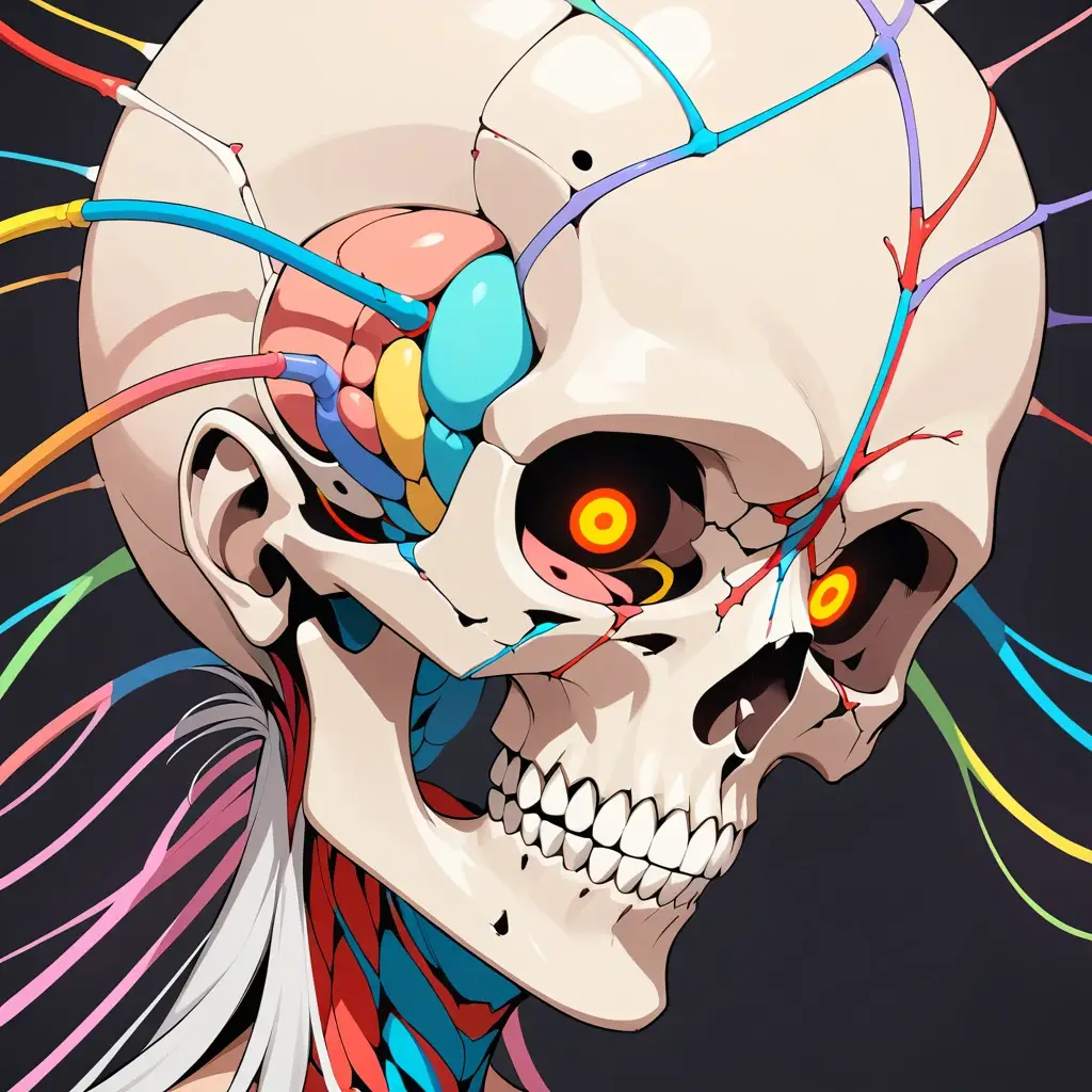
The human skull is a complex, protective structure housing the brain, sensory organs, and essential passageways for nerves and vessels. But what exactly is the difference between the cranium and the skull? Which bones form the neurocranium and viscerocranium? How do sutures close, and how do ossification timelines vary? This comprehensive guide breaks it all down—from bone-by-bone analysis to clinical relevance like epidural hematomas and craniosynostosis. For a quick intro, check out our explainer on where the cranial is.
Understanding Terminology
What is the Skull?
The skull includes all 22 bones of the head—both the cranial and facial bones. It protects the brain and supports the face.
What is the Cranium?
The cranium is the portion of the skull that encloses the brain—excluding the facial bones. It’s often used interchangeably with “skull,” but medically, it refers specifically to the neurocranium.
Cranial vs. Facial Skeleton
Neurocranium
This forms the protective case around the brain and consists of 8 bones.
Calvaria (Skullcap)
Includes the frontal, parietal, and upper occipital bones forming the roof of the cranium.
Cranial Base & Fossae
The base includes the ethmoid, parts of the sphenoid, and occipital bones. Fossae are the anterior, middle, and posterior depressions that cradle the brain.
Viscerocranium (Facial Skeleton)
The viscerocranium forms the framework of the face. It supports eating, breathing, and communication.
Detailed Overview of Cranial Bones
Frontal Bone
Forms the forehead and upper part of the eye sockets. Contains the frontal sinuses.
Parietal Bones
Paired bones forming the sides and roof of the cranium. Join at the sagittal suture.
Temporal Bones
House structures of the ears and include key foramina like the carotid canal.
Occipital Bone
It forms the back of the skull and contains the foramen magnum, where the spinal cord exits.
Sphenoid and Ethmoid Bones
- Sphenoid: Central bone with multiple foramina for nerves (optic canal, foramen ovale, spinosum).
- Ethmoid: Forms the nasal septum and contains the cribriform plate for olfactory nerves.
Facial Bones Breakdown
Mandible & Maxilla
- Mandible: Lower jaw, forms the temporomandibular joint (TMJ).
- Maxilla: Upper jaw, forms part of the orbit and nasal cavity.
Zygomatic, Nasal, Lacrimal, Palatine
Support facial structure, house the tear ducts, and form part of the eye socket.
Inferior Nasal Conchae and Vomer
Shape the nasal cavity and help filter and humidify inhaled air.
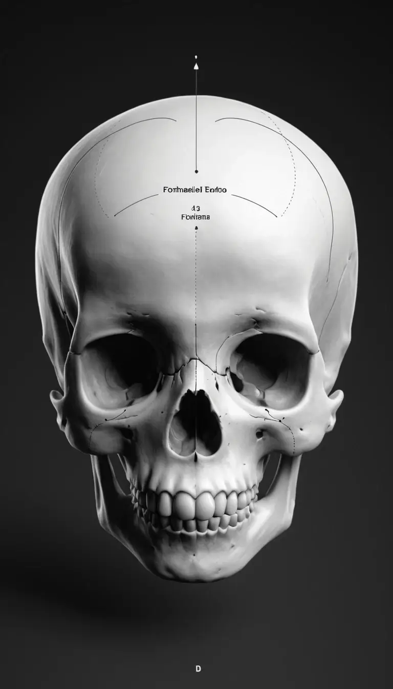
Cranial Sutures & Fontanelles
The cranial bones are joined by sutures (immovable joints), including:
- Coronal
- Sagittal
- Lambdoid
- Squamous
In infants, these bones are separated by fontanelles. Key closure times:
- Anterior fontanelle: ~18 months
- Posterior fontanelle: ~2-3 months
This aligns with popular search queries like “suture closure age by bone.”
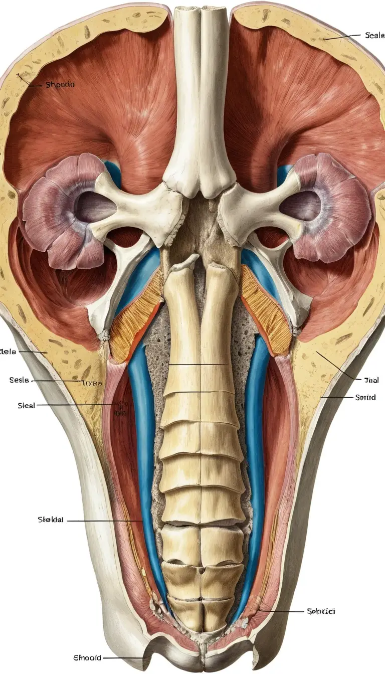
Important Foramina & Neurovascular Passages
Foramina allow passage of nerves and vessels.
- Optic canal: CN II (optic nerve)
- Foramen rotundum: CN V2
- Jugular foramen: CN IX, X, XI, and jugular vein
- Foramen magnum: Brainstem to spinal cord
A full cranial nerve foramina list with diagrams enhances value for students and clinicians.
Development & Embryology of Skull Bones
Skull bones develop via:
- Intramembranous ossification (flat bones like parietal and frontal)
- Endochondral ossification (base of skull)
Cranial Ossification Timeline
- Begins in utero (~week 8)
- Fontanelles ossify postnatally
- Sutures fuse in early adulthood (varies by bone)
Differences in adult vs. fetal skull anatomy are critical in pediatrics and embryology.

Common Clinical Conditions
Craniosynostosis
Premature fusion of one or more sutures. Leads to skull deformities and possible brain pressure.
Epidural Hematoma & Pterion Anatomy
The pterion is a thin area where four bones meet. A blow here can rupture the middle meningeal artery, causing a fast-bleeding epidural hematoma—a medical emergency.
Facial Fractures & Orbital Blowout
Common in trauma. The orbital floor is particularly vulnerable, potentially trapping eye muscles.
Frequently Confused Terms
- Cranium vs Skull: Cranium = braincase; Skull = cranium + facial bones
- Calvaria vs Cranial Base: Calvaria is the dome; base is the floor of the cranial cavity
- Neurocranium vs Viscerocranium: Braincase vs. Facial Bones
Why the Distinction Matters
Clear anatomical terminology improves communication in:
- Medical diagnosis
- Surgery
- Anatomy education
It helps avoid life-threatening mistakes—especially in trauma or pediatric cases.
Summary & Key Takeaways
- The skull is divided into neurocranium and viscerocranium
- Development involves different ossification processes
- Key clinical zones include the pterion and cranial sutures
- Understanding terms like “calvaria,” “fontanelles,” and “cranial fossae” boosts academic and clinical precision
Suggested External Resources
For deeper learning and credible references:
- Wikipedia: Human Skull
- Forbes Health—on cranial conditions
- Radiopaedia—great for imaging and clinical correlations
Leave a Reply