 Introduction to Cranial Anatomy
Our skull isn’t just a bone shell protecting the brain—it’s a highly structured, multifunctional masterpiece that plays a major role in both protection and function. From cranial nerve diagrams to understanding disorders like cranial nerve palsy, there’s so much more than meets the eye when we talk about the “cranial” system.
 Definition of “Cranial”
Cranial refers to anything related to the skull, particularly the part that encloses the brain. It’s a key term in anatomy and is often used when discussing head injuries, neurological assessments, and medical imaging.
 Origin and Etymology of the Term
The term cranial comes from the Greek word kranion, meaning skull. Over time, it has evolved into a modern medical term with significant usage in both clinical and academic settings.
 Synonyms & Related Terms
You may come across cephalic and intracranial used interchangeably with cranial in medical literature. While all relate to the head, “cephalic” often includes the entire head region, and “intracranial” specifically refers to the space within the skull.
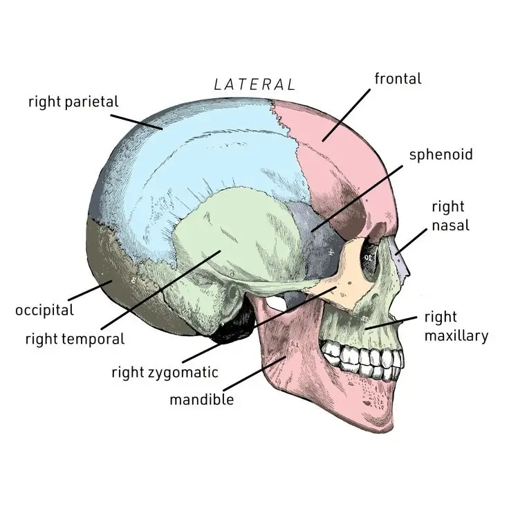
 Cranial Bones and Structure
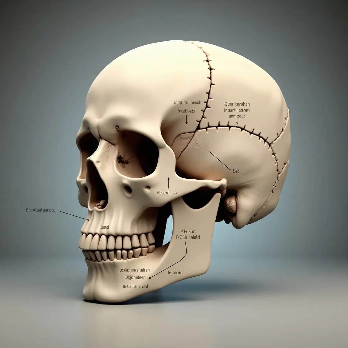
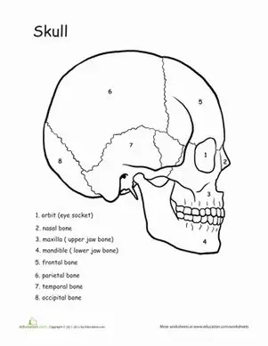
Overview of Cranial Bones
The cranium is composed of eight primary bones, including the frontal, parietal, temporal, occipital, sphenoid, and ethmoid bones. These bones fuse together in adulthood and create a protective case around the brain.
Major Cranial Bones Explained
Frontal Bone: Forehead and upper eye sockets
Parietal Bones: Upper sides and roof of the skull
Occipital Bone: Back and base of the skull
Temporal Bones: Lower sides near the ears
Functions of the Cranium
The skull isn’t just there for protection—it also gives shape to your face, supports sensory structures, and provides attachment points for muscles.
 Cranial Capacity and Its Measurement
Methods of Measurement
Cranial capacity can be measured using MRI or CT volumetric imaging or by using traditional anthropometric methods like the cranial index.
Relevance in Neurology and Anthropology
A larger cranial capacity often correlates with greater brain volume, which is significant in both neurological studies and evolutionary anthropology.
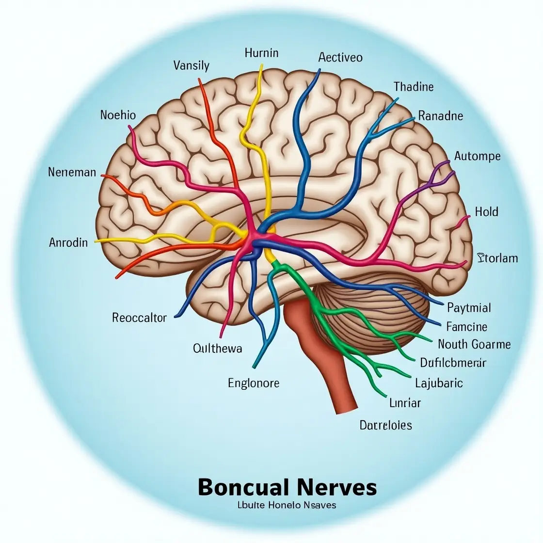
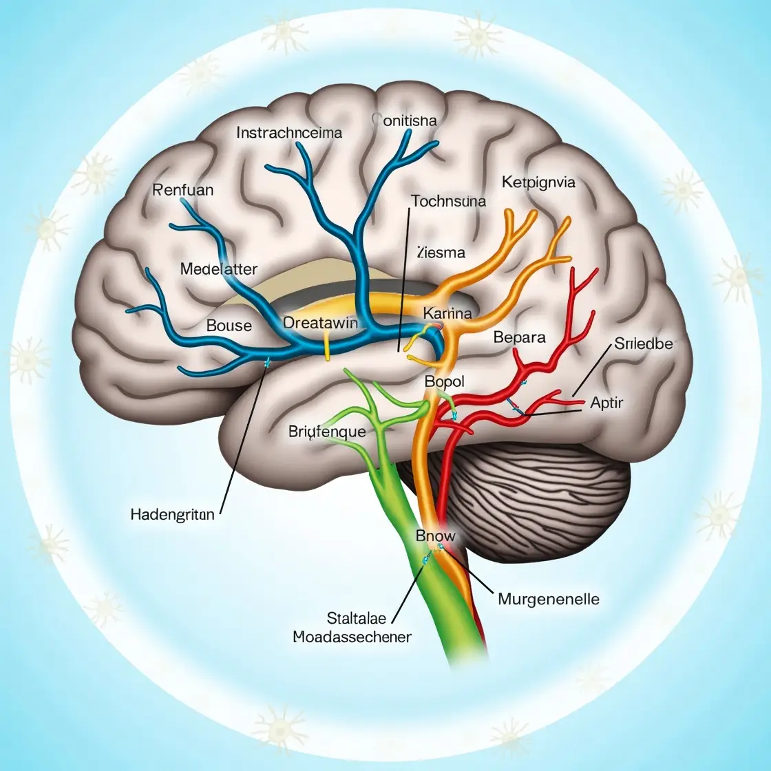
Understanding Cranial Nerves
What Are Cranial Nerves?
There are 12 cranial nerves, and each has a specific function, such as moving muscles, sensing smells, or controlling heart rate.
List of the 12 Cranial Nerves
Some key ones include
Olfactory (I): Smell
Optic (II): Vision
Facial (VII): Facial expressions
Vagus (X): Control of internal organs
Cranial Nerve Functions in Detail
Sensory vs. Motor Functions
Some nerves, like the optic nerve, are purely sensory, while others, like the trochlear, are purely motor. A few, like the vagus nerve, are mixed.
Common Pathways
Cranial nerves travel through foramina (holes) in the skull, making them vulnerable to trauma or surgical damage.
 Disorders Affecting the Cranial System
Cranial Nerve Palsy
Affects motor control of muscles, especially the eyes and face. Common types include Bell’s palsy and sixth nerve palsy.
Treatment Options
These range from corticosteroids and antivirals to physical therapy.
Cranial Nerve Damage After Surgery
It often occurs after procedures near the base of the skull. Recovery time varies, typically between weeks and months, depending on nerve regeneration.
Intracranial Pressure: Symptoms and Monitoring
Symptoms include:
Headache
Blurred vision
Vomiting
Tools like intracranial pressure monitoring devices help in emergency neurology.
Cranial Nerve Disorders: Symptoms and Diagnosis
Common symptoms:
Numbness
Paralysis
Pain
Diagnosis includes MRI, CT, and EMG tests.
 Diagnostic and Imaging Techniques
MRI & CT Scans for the Cranium
High-resolution scans help visualize internal bone structure and detect abnormalities.
Electromyography (EMG) for Cranial Nerves
Used to assess the electrical activity of cranial muscles and diagnose nerve dysfunctions.
 Medical Terminology & Usage
Terms like “cranial cavity,” “cranial index,” and “cranial nerves” are frequently used in medical records and textbooks (source: Wikipedia).
Comparative Cranial Anatomy
How Cranial Structures Differ Across Species
Humans have more rounded cranial cavities compared to quadrupeds, which helps accommodate a larger brain.
Evolutionary Significance of the Cranium
Skull evolution has been pivotal in the development of higher brain functions in mammals, especially primates.
 Conclusion
Understanding cranial anatomy is more than just memorizing bones—it’s about appreciating a complex system that protects, supports, and connects vital neural functions.
For an in-depth look at cranial bones and health, check out our detailed guide here: The Ultimate Guide to Cranial Bones: Anatomy, Functions, and Health Tips
 FAQs
FAQs
1. What are the most common cranial nerve disorders?
Bell’s palsy, trigeminal neuralgia, and optic neuritis are among the most frequently reported.
2. How is cranial capacity measured in modern medicine?
Via MRI volumetric imaging or using cranial indices derived from caliper measurements.
3. What are signs of elevated intracranial pressure?
Severe headache, nausea, and blurred vision are early signs.
4. Can cranial nerve damage heal naturally?
Yes, depending on the severity. Some nerves regenerate, especially if damage is minor.
5. What is the difference between cranial and cephalic?
“Cranial” refers specifically to the skull; “cephalic” refers to the entire head region.
Leave a Reply